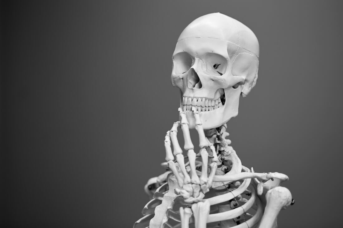
 Mathew Schwartz Mathew Schwartz Whether it’s on television, in movies or in real life, have you ever noticed how someone reacts to getting hit in the mouth or face? In most cases they will move their lower jaw from side to side, stretch their mouth open, slam it shut, and generally try to put their face back the way it should be. While most people usually haven’t been hit in the mouth, those who have temporomandibular joint (TMJ) dysfunction certainly can empathize with these unfortunate individuals. When the temporomandibular joint is not in place and working properly it can cause a host of serious problems, such as grinding or “popping” of the jaw, an occasional unhinging or dislocation sensation when one opens their mouth, or just plain constant chronic soreness around the back teeth and ears. TMJ dysfunction affects everyone of all ages. There are many causes of TMJ dysfunction. They include stress, dental malocclusion (imperfect bite) and even arthritis. Often poor posture is to blame especially in the alignment of one’s neck bones and how one holds their head. One point to remember is; even though you have two separate temporomandibular joints, they must move simultaneously or at least in harmony. When proper movement is impaired, pain develops. To understand the situation better, let’s look exactly where all this occurs. The TMJ is the joining of the inferior maxillary (jaw) bone with each temporal bone on either side of the base of the cranium. The skull consists of 22 bones, 14 cranial and 8 facial. All are fused together into a single unit, save for one. Of course that one bone is the jaw bone. Hence, the need for a joint to hold this bone to the rest of the skull. A joint with ligaments, membranes, cartilage and the potential for all sorts of trouble. Like all other joints (such as the knee, wrist and ankle) the TMJ is extremely vulnerable to strains, inflammations and everyday wear and tear (especially when aggravated by poor posture). The TMJ is a form of a ball and socket joint, with the condyle (knobby end) of the jaw bone and the glenoid cavity of the temporal bone representing the ball and socket, respectively. It is interesting to note that the angle of the condyle (and the neck) changes as we develop and grow older. The bones are held together by a group of five ligaments: the external lateral, internal lateral, stylo-mandibular, capsular and interarticular fibro-cartilage. The two lateral ligaments attach on either side of the jawbone (inside and outside). The capsular ligament attaches to the condyle of the jawbone, while the stylo-mandibular ligament attaches to the angle of the jaw (at the bottom-rear, where one feels the soreness of the jaw). The interarticular fibro-cartilage, sandwiched between and in conjunction with two synovial membranes serves as a cushion between the bones. The TMJ’s configuration allows the extensive range of motion one experiences with their jaw bone. Like the body itself, however, what we take for granted is the result of an extremely complex structure. It’s this complexity that leaves the individual susceptible to the different cause of discomfort we described before. The temporomandibular joint is classified as a hinge-type joint, but it has a much more complex action than that. When simply opening and closing the mouth, the TMJ acts as a hinge, However, in the chewing action there is a complex movement of the joint which provides the grinding action of the teeth. It is impossible for only one side of the jaw to move at a time. During the grinding action, one temporomandibular joint slides forward while the other slides back. you can feel this by placing your fingers on your jaw joints and moving your jaw to the side (as if chewing). While your fingers are in this position, you can observe for “clicking” of the jaw. There should be no clicking or popping of the joint as the TMJ moves through its complete range of motion. Sometimes this noise is audible to people close to you; other times it can only be felt as a lack of smooth movement and heard only by you. Popping and clicking of the TMJ indicates that it is not functioning normally. Examination includes determining the balance of TMJ activity and the muscles which move the jaw through its range of motion. When an imbalance is found, it can often be corrected by balancing the jaw’s muscular activity with physical therapy techniques. These treatments usually produce an immediate balancing of the muscles. It is sometimes necessary to have the bite balanced by a dentist to maintain muscle and TMJ joint balance. The medical team of Dentists and Physical Therapists have long known that TMJ dysfunction can cause symptoms far removed from the joint itself. Headaches, back pain, and pain across the shoulders are often relieved after TMJ dysfunction is corrected. More recent evidence shows that the TMJ dysfunction can cause functional problems throughout the body. Some of the more common symptoms of a TMJ dysfunction may include: facial pain, facial muscle fatigue or spasms, headaches, pain and/or “popping or clicking” of the jaw (especially while eating), tenderness or inflammation of the joint, dislocation or even locking of the jaw. Any one of these symptoms may indicate a serious problem, and it is recommended that the person having these symptoms see their dentist and have him refer that person for treatment. It is estimated that between 5 and 20 percent of the population suffer from temporomandibular disorders. Until recently, most of these patients, the vast majority of whom are female, have been labeled as at least partly psychotic. However, research efforts in the past 40 years and more have clearly shown that psychoses probably play a minor role in TMJ disorders and that stress may be the dominant factor in the etiology of the specific problem. The importance of referred pain cannot be overestimated in this disorder. As with any other problem, proper diagnosis is the essential key to successful treatment. Don’t put it off! If you would like more specific information on TMJ, please feel free to contact my office at 626-576-0591
0 Comments
 Image by Alexas_Fotos from Pixabay Image by Alexas_Fotos from Pixabay Things you can do to help yourself until your physician can see you When tissue is damaged by being crushed, stretched, or torn, an inflammatory response occurs. Hemorrhage and swelling occur, along with the direct effect of the inflammatory products on nerve endings, produce pain and spasm. The spasm not only splints the damaged tissue, but also compresses pain fibers. Moreover, it lessens the efficiency of the vascular system and can result in ischemia (lack of blood supply) at the injured area. The pain fiber compression and ischemia establish a vicious pain spasm cycle. As a result, more cells are eventually injured than were initial. A hematoma forms when blood vessels are torn. Plasma also leaks into the damaged area to produce edema (swelling). Swelling is the greatest enemy of healing. The goal of early treatment is to delay or minimize swelling. By limiting the swelling, pain and muscle spasm decrease and the magnitude of the injury is reduced. Early motion is also important; otherwise, if the injured person avoids using the part, his/her muscles will atrophy in a few days. The joint or damaged tissue will then be less stable and will most likely be easy to injure again. ICE: The appropriate first-aid treatment for soft-tissue injuries is the application of ice. Ice, by delaying or minimizing swelling, decreases pain and muscle spasm and limits the magnitude of an injury. The lower tissue temperature induces local vein constriction, which lessens capillary permeability, making the blood more viscous or thick. Less blood flows into the injured area, and the hematoma (or local swelling) is less. The ice dulls peripheral pain by interfering locally with nerve impulses and decreasing the nerve’s ability to transfer signals. It relieves spasms by decreasing muscle activity, muscle spindle firing, and restricts certain chemicals which inhibit blood flow. Ice limits the magnitude of an injury by lowering metabolism in peripheral, uninjured cells. By decreasing the cellular demand for oxygen, cells are put in partial hibernation, and thus extension of the injury is avoided. Otherwise cells that have survived the initial trauma may not be able to withstand the lack of oxygen imposed by the disruption of local circulation. Initial treatment of an injury follows the acronym ICE, meaning Ice, Compression, and Elevation. The steps to be taken are as follows:
Heat: Over the years, heat in various forms has been the standard therapeutic treatment. Cold is now being applied more frequently, not only for the initial treatment of the acute injury, but also for the recovery phase. Heat is used now only in rehabilitation, whereas ice may be used to treat acute injuries and in rehabilitation. Some people are averse and sensitive to cold. The choice of heat for rehabilitation, then may be based on the injured person’s comfort. Both ice and heat are analgesic and reduce muscle spasm, but in most cases heat produces more comfort as a result of its sedating properties. Heat induces vein dilation and increases blood flow, resulting in an influx of oxygen and nutrients to the injured area and waste products being carried away. Regardless of whether heat or cold is used, however it still takes time for an injury to heal. After acute symptoms have subsided, the recovery rate with heat or cold is about the same. Waterproof heating pads are especially good for home first-aid. A moist towel should be placed between the pad and the skin to provide moist heat. The objectives of rehabilitation are to regain range of motion, strength, flexibility, muscular endurance, power, cardiovascular endurance, speed, balance agility and skills. The rehab process comprises many steps and each one must be successfully completed pain-free. In all cases of severe injury, consult your physician for a professional examination immediately! |
Sheila’s BlogI focus on the topics you care about most. Categories
All
Archives
February 2022
|
|
55 S. Raymond Ave. Suite 100
Alhambra, CA 91801 Main Phone: (626) 576-0591 Alternate Phone: (626) 538-3966 Fax: (626) 576-5890 Email: yonemotoptfinance@gmail.com |
© 2015 Yonemoto Physical Therapy. ALL RIGHTS RESERVED.
|


 RSS Feed